Testicular
Cancer Overview
 Testicular
cancers are relatively rare but highly curable, and occur predominantly
in young and middle aged males. Testicular cancers were among the first
types of cancers to be cured by radiation and/or chemotherapy, and
treatment has been refined over the last two decades. Currently, over
70% of all patients are curable regardless of the extent of cancer.
Thus, all treatment of testicular cancer is delivered with the intent
to cure. However, it is important to know the extent of cancer and the
specific type of testicular cancer in order to administer the best
therapy.
Testicular
cancers are relatively rare but highly curable, and occur predominantly
in young and middle aged males. Testicular cancers were among the first
types of cancers to be cured by radiation and/or chemotherapy, and
treatment has been refined over the last two decades. Currently, over
70% of all patients are curable regardless of the extent of cancer.
Thus, all treatment of testicular cancer is delivered with the intent
to cure. However, it is important to know the extent of cancer and the
specific type of testicular cancer in order to administer the best
therapy.
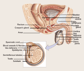
The testicles are located inside the scrotum (a
sac of loose skin that lies directly under the penis). The testicles
are similar to the ovaries in women. Sperm and male hormones are made
in the testicles. Testicular cancer—also called germ cell cancer—occurs
in the tissues of one or both testicles. Similar cancers called
"non-gonadal germ cell cancers" can also occur outside the testicle;
non-gonadal germ cell cancers are not discussed in this section.
Testicular cancer is the most common cancer in men
15 to 35 years old. Men who have an undescended testicle (a testicle
that has never moved down into the scrotum) are at higher risk of
developing testicular cancer than men whose testicles have moved
normally down into the scrotum. This is true even if surgery has been
performed early in life to place the testicle in the appropriate place
in the scrotum.
A swelling in the scrotum is usually the first
sign of testicular cancer. A doctor will examine the testicles to feel
for any lumps. If any lumps are found, the doctor will perform an
ultrasound examination, which uses sound waves to make a picture of the
inside of the testes. In addition, the physician may perform a computed
tomography (CT) or positron emission tomography (PET) scan to determine
whether cancer is present. A PET scan is similar to a CT scan; however,
PET scans can detect live cancer tissue. Prior to a PET scan, the
patient receives an injection of a substance that contains a type of
sugar attached to a radioactive isotope. The cancer cells “take up” the
sugar and attached isotope, which emits positively charged, low energy
radiation (positrons). The positrons react with electrons in the cancer
cells, which creates the production of gamma rays. The gamma rays are
then detected by the PET machine, which transforms the information into
a picture. If no gamma rays are detected in the scanned area, it is
unlikely that the mass in question contains living cancer cells.
When cancer is suspected, the entire testicle is
surgically removed (orchiectomy) through an incision in the groin. The
surgically removed tissue is then examined under a microscope to
determine whether cancer cells are present. Removal of a small piece of
tissue (biopsy) is usually not done because this is thought to cause
spread of the cancer. When the cancer is small and localized to the
testicle, removal of the testicle may be all of the treatment that is
necessary to cure the cancer. The surgically removed testicle is
examined under the microscope to determine the type of cancer. In some
patients the cancer consists of only one cell type. But for many
patients, the cancer under the microscope consists of a mixture of cell
types.
Testicular cancer is broadly divided into two
different types, seminoma and nonseminoma, based on the appearance of
cells under the microscope. Nonseminomas are, in general, more
difficult to cure than seminomas. Nonseminoma cell types include:
embryonal carcinoma, teratoma, yolk sac carcinoma, choriocarcinoma, and
various combinations that are referred to as “mixed cell types”. For
nonseminoma cancer teratoma presents the lowest risk of spread and
choriocarcinoma presents the highest risk of spread; the other cell
types are of intermediate risk.
Treatment planning depends upon whether the
testicular cancer is classified as seminoma or nonseminoma. Seminomas
are more sensitive to radiation therapy and are easier to cure than
nonseminomas. Patients with all stages of seminoma have a cure rate
that exceeds 90%, and patients with seminoma confined to the testicle
have a cure rate approaching 100%. If there is a mixture of seminoma
and nonseminoma components upon examination under the microscope, the
cancer is diagnosed as nonseminoma because the cancer will be more
aggressive due to the nonseminoma part of the cancer.
The extent of disease, or “stage” is determined
after surgical removal of the testicle. All patients will require CT or
magnetic resonance imaging (MRI) scans of the abdomen, chest, and
sometimes the brain or bones to look for spread of disease beyond the
testicle.
Lymph nodes are small, bean-shaped structures that
are an essential component of the immune system. They are found
throughout the body and are interconnected with lymph channels.
Testicular cancer tends to spread through lymph channels that drain
into lymph nodes in the groin area, into channels near the large blood
vessel (the aorta) carrying blood from the heart, and into lymph nodes
between the abdomen and back called retroperitoneal lymph nodes.
Tumor or Cancer Markers
An important aspect of the evaluation of testicular cancer is the use
of blood or serum tests to detect cancer markers. Cancer markers are
abnormal substances in the blood associated with the presence of cancer
somewhere in the body. Common cancer markers that are present in the
blood of patients with testicular cancer include:
- Alpha-fetoprotein (AFP)
- Beta human chorionic gonadotropin (beta-hCG)
- Lactate dehydrogenase (LDH)
These cancer markers may detect cancers that are
too small to be detected with a CT scan. In males under age 15, about
90% of testicular germ cell cancers are yolk sac tumors that make AFP,
which is an excellent indicator of response to therapy and disease
status.
It is important to realize that the absence of
cancer markers in the blood following treatment does not always mean
the absence of cancer, even when cancer markers were present at
diagnosis. Patients who appear to have seminoma when the cancer is
examined under the microscope and have elevated serum levels of AFP are
treated as if they have nonseminoma because seminoma cells do not
secrete this cancer marker and other cell types must be present, even
though they may not be visible under the microscope. Elevation of the
beta-hCG is found in approximately 10% of patients with pure seminoma
and
is an indication of metastatic spread of disease, but does not change
the cellular diagnosis.
Type of treatment and outcomes depend on the stage
and spread of the cancer. In order to learn more about the most recent
information available concerning the treatment of testicular cancer,
click on the appropriate stage.
NonSeminoma - Stages & Types of Treatment |
|
Reference:
--------------------------------------------------------------------------------
[1] Jewett MAS, Groll RJ. Nerve-sparing
retroperitoneal lymphadenectomy. Urologic Clinics of North America.
2007;34:149-158.
2. Syndication Cancer Consultants.